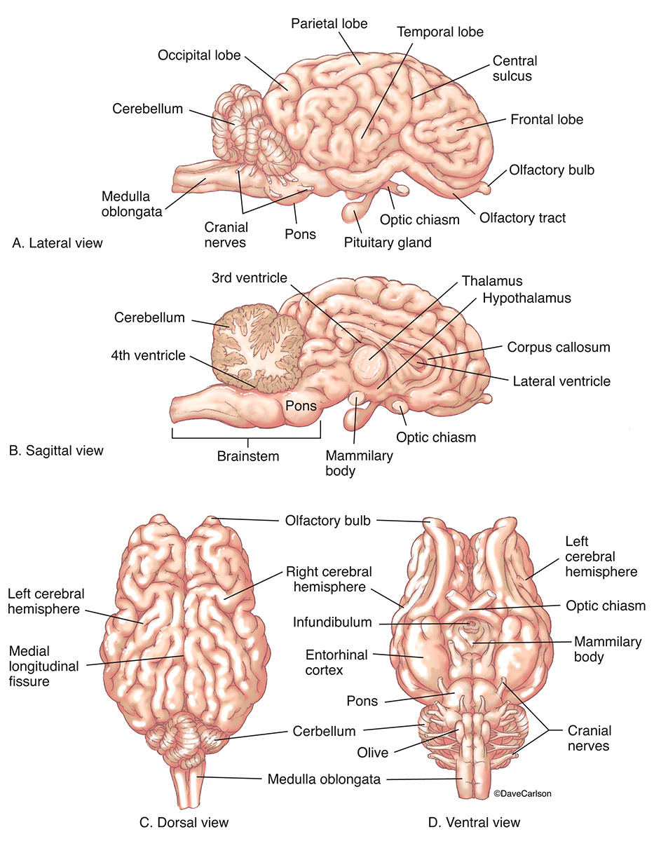
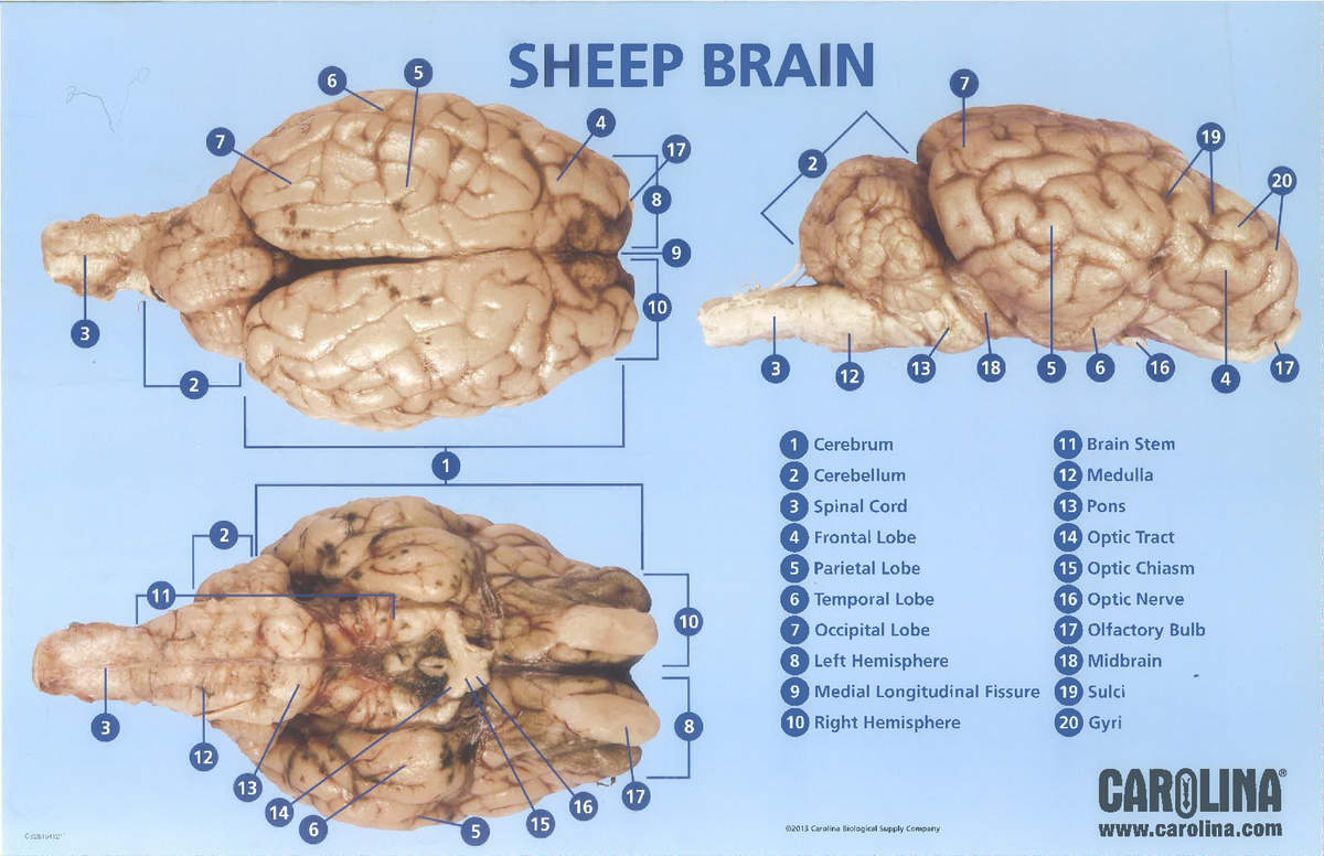
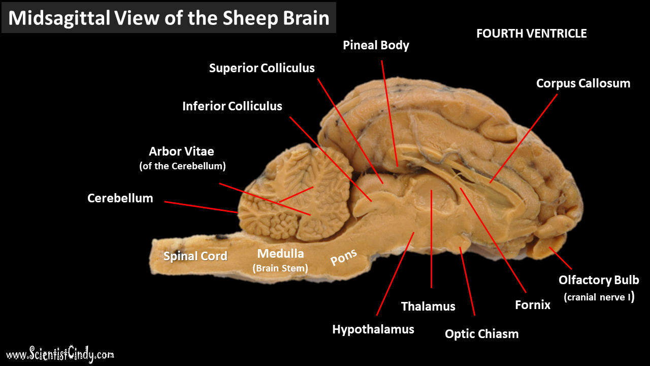
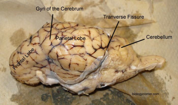
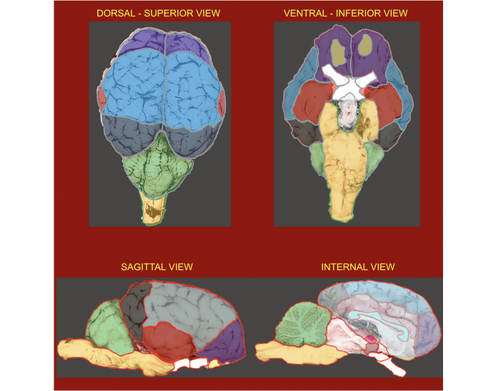
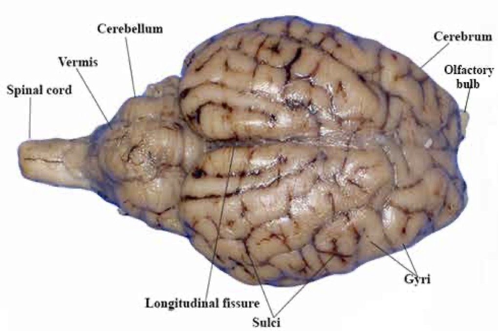
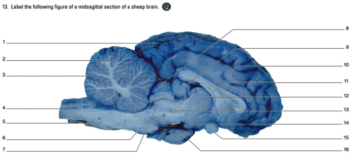
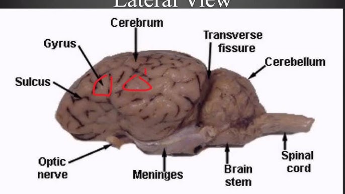
38 sheep brain diagram labeled
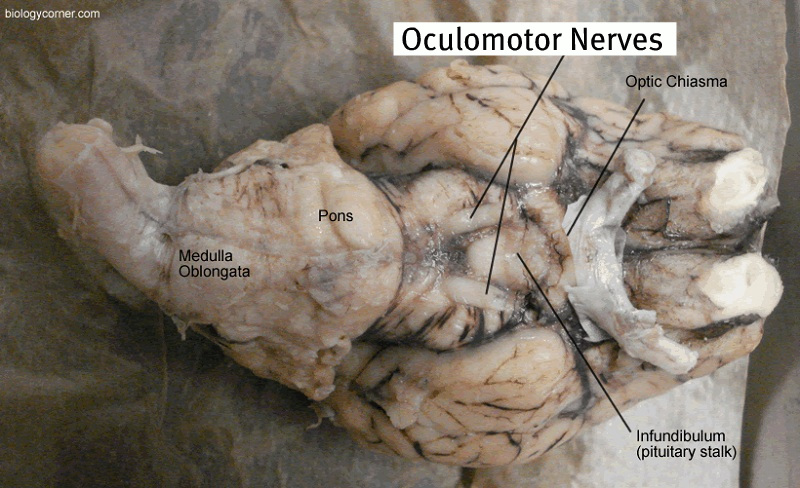
bio.libretexts.org › Bookshelves › Human_Biology11.7: Sheep Brain Dissection - Biology LibreTexts The sheep brain is enclosed in a tough outer covering called the dura mater. You can still see some structures on the brain before you remove the dura mater. Take special note of the pituitary gland and the optic chiasma. These two structures will likely be pulled off when you remove the dura mater. Figure 11.7. 1: Brain with Dura Mater Intact › anatomy › sheepbrainSheep Brain Label - The Biology Corner Label theBrain of the Sheep Publisher: Biologycorner.com ; follow on Google+ This work is licensed under a Creative Commons Attribution-NonCommercial 3.0 Unported License .
learning-center.homesciencetools.com › article › brain-dissection-projectSheep Brain Dissection Project Guide | HST Learning Center Sheep Brain Dissection: Internal Anatomy Place the brain with the curved top side of the cerebrum facing up. Use a scalpel (or sharp, thin knife) to slice through the brain along the center line, starting at the cerebrum and going down through the cerebellum, spinal cord, medulla, and pons.

Sheep brain diagram labeled
quizlet.com › 264003651 › sheep-brain-dissection-labeled-diagramSheep Brain Dissection labeled Diagram | Quizlet Sheep Brain Dissection labeled Diagram | Quizlet Sheep Brain Dissection labeled + − Learn Test Match Created by AllieKlinger Terms in this set (8) Corpus Collosum ... Lateral Ventricle ... Fornix ... Hypothalamus ... Cerebral Aqueduct ... Central Canal ... Inferior Collicuious ... Transverse Fissure ... sydneypfleiger › teacher-resources › InteractiveSheep Brain Dissection | Carolina.com Sheep Brain Dissection. The body consists of organ systems, each with specific roles for maintaining life. Each organ system consists of organs that operate together to carry out certain body functions. Organ systems do not operate independently but are coordinated for the organism to survive. › arts › psychologySheep Brain Neuroanatomy Online Self-Test | KPU.ca - Kwantlen ... Sheep Brain Neuroanatomy Online Self-Test Use each diagram as a reference, and selected the correct answer for each lettered structure. You may find it useful to open the diagrams in a separate window to review while answering each question. Dorsal Surface Dorsal Surface A * Occipital Lobe Temporal Lobe Cerebellum Parietal Lobe Dorsal Surface B *
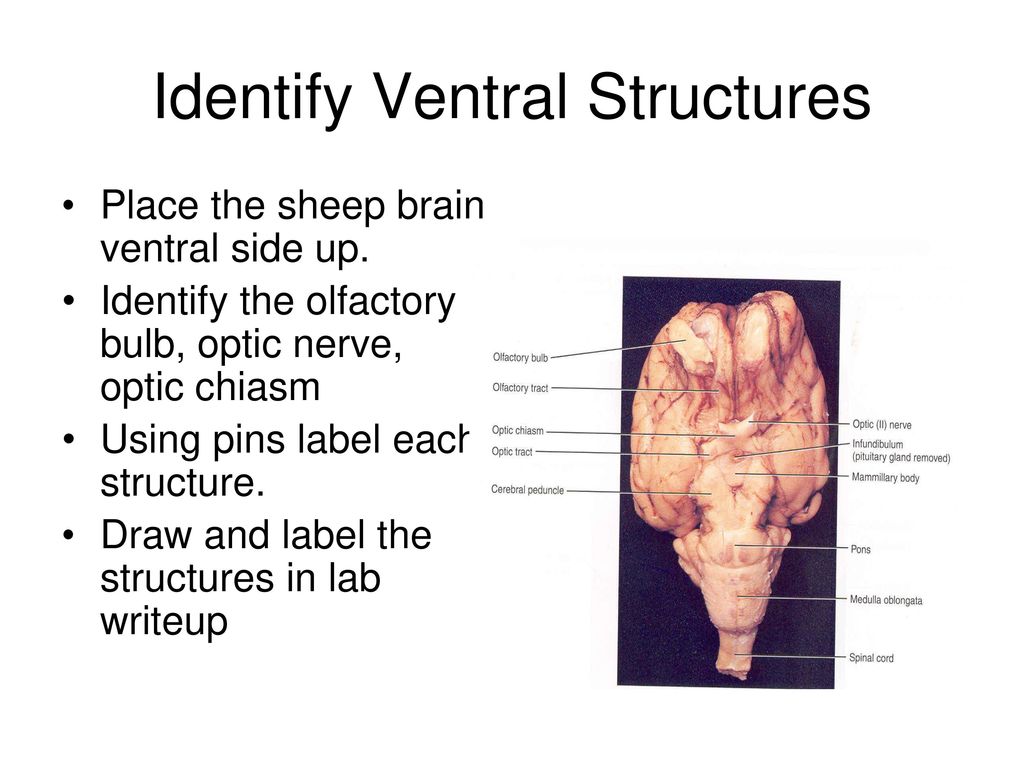
Sheep brain diagram labeled. quizlet.com › 436514897 › sheep-brain-diagramSheep Brain Diagram | Quizlet Optic nerve the nerve that carries neural impulses from the eye to the brain Olfactory bulb the brain center for smell, located below the frontal lobes Optic chiasm the point in the brain where the visual field information from each eye "crosses over" to the appropriate side of the brain for processing Medulla oblongata › anatomy › sheepbrainSheep Brain Dissection with Labeled Images - The Biology Corner Sheep Brain Dissection with Labeled Images Sheep Brain Dissection 1. The sheep brain is enclosed in a tough outer covering called the dura mater. You can still see some structures on the brain before you remove the dura mater. Take special note of the pituitary gland and the optic chiasma. learning-center.homesciencetools.com › wp-content › uploadsSheep Brain Dissection Lab - Home Science Tools Print out the diagrams on the following pages and fill in the labels to test your knowledge of sheep brain anatomy. External anatomy: label the top view (.pdf) External anatomy: label the bottom view (.pdf) Internal anatomy: label the right side (.pdf) See our other free dissection guides with photos and printable PDFs. Click here. anatomylearner.com › sheep-brain-anatomySheep Brain Anatomy with Labeled Diagram Nov 16, 2022 · The sheep brain anatomy consists of 3 major parts – prosencephalon (forebrain), mesencephalon (midbrain), and rhombencephalon (hindbrain). These 3 main parts of the sheep brain again divide into specific segments. There are also 5 different lobes in the sheep brain structure – frontal, parietal, occipital, temporal, and limbic area.
› arts › psychologySheep Brain Neuroanatomy Online Self-Test | KPU.ca - Kwantlen ... Sheep Brain Neuroanatomy Online Self-Test Use each diagram as a reference, and selected the correct answer for each lettered structure. You may find it useful to open the diagrams in a separate window to review while answering each question. Dorsal Surface Dorsal Surface A * Occipital Lobe Temporal Lobe Cerebellum Parietal Lobe Dorsal Surface B * › teacher-resources › InteractiveSheep Brain Dissection | Carolina.com Sheep Brain Dissection. The body consists of organ systems, each with specific roles for maintaining life. Each organ system consists of organs that operate together to carry out certain body functions. Organ systems do not operate independently but are coordinated for the organism to survive. quizlet.com › 264003651 › sheep-brain-dissection-labeled-diagramSheep Brain Dissection labeled Diagram | Quizlet Sheep Brain Dissection labeled Diagram | Quizlet Sheep Brain Dissection labeled + − Learn Test Match Created by AllieKlinger Terms in this set (8) Corpus Collosum ... Lateral Ventricle ... Fornix ... Hypothalamus ... Cerebral Aqueduct ... Central Canal ... Inferior Collicuious ... Transverse Fissure ... sydneypfleiger











Post a Comment for "38 sheep brain diagram labeled"