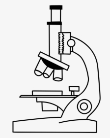
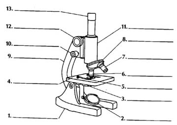
40 light microscope drawing with label
› publication › ppic-statewide-surveyPPIC Statewide Survey: Californians and Their Government Oct 26, 2022 · Key Findings. California voters have now received their mail ballots, and the November 8 general election has entered its final stage. Amid rising prices and economic uncertainty—as well as deep partisan divisions over social and political issues—Californians are processing a great deal of information to help them choose state constitutional officers and state legislators and to make ... Compound Microscope Parts - Labeled Diagram and their Functions Labeled diagram of a compound microscope Major structural parts of a compound microscope Optical components of a compound microscope Eyepiece Eyepiece tube Objective lenses Nosepiece Specimen stage Coarse and fine focus knobs Rack stop Illuminator Condenser Abbe condenser Iris Diaphragm Condenser Focus Knob Summary An overview of microscopes
Microscope Drawing: How to Sketch Microscope Slides How to Draw Microscope Slides Organize and orient your field of view: To begin, draw a circle as large as possible with a pencil. An 8.5 x 11-inch piece of paper is good size for beginners. The circle represents what you see through the eyepiece of the microscope. Using thin lines, divide the circle into quarters in order to organize the picture.
Light microscope drawing with label
› cells › cell_modelInteractive Eukaryotic Cell Model - CELLS alive Cytosol: The cytosol is the "soup" within which all the other cell organelles reside and where most of the cellular metabolism occurs.Though mostly water, the cytosol is full of proteins that control cell metabolism including signal transduction pathways, glycolysis, intracellular receptors, and transcription factors. Label the microscope — Science Learning Hub Label the microscope Interactive Add to collection Use this interactive to identify and label the main parts of a microscope. Drag and drop the text labels onto the microscope diagram. eye piece lens diaphragm or iris coarse focus adjustment stage base fine focus adjustment light source high-power objective Download Exercise Tweet Givenchy official site WebDiscover all the collections by Givenchy for women, men & kids and browse the maison's history and heritage
Light microscope drawing with label. Plant Cell Under Microscope Drawing To use a light microscope to examine animal or plant cells. Plant cell under microscope drawing. Drawings MUST be completed neatly using a pencilcolored pencil. Students will finish plant cell diagrams from Monday. Onion Cell drawing high power 2. A cell is a very tiny structure which exists in living bodies. Interactive Eukaryotic Cell Model - CELLS alive WebSecretory Vesicle: Cell secretions - e.g. hormones, neurotransmitters - are packaged in secretory vesicles at the Golgi apparatus.The secretory vesicles are then transported to the cell surface for release. Cell Membrane: Every cell is enclosed in a membrane, a double layer of phospholipids (lipid bilayer).The exposed heads of the bilayer are "hydrophilic" … Parts of a microscope with functions and labeled diagram - Microbe Notes Parts of a microscope with functions and labeled diagram September 17, 2022 by Faith Mokobi Having been constructed in the 16th Century, Microscopes have revolutionalized science with their ability to magnify small objects such as microbial cells, producing images with definitive structures that are identifiable and characterizable. › createJoin LiveJournal Password requirements: 6 to 30 characters long; ASCII characters only (characters found on a standard US keyboard); must contain at least 4 different symbols;
Light Microscope: Functions, Parts and How to Use It To use a light microscope, you can follow the steps below carefully. Start with a low lens and a clean slide. The microscope stage should be lowered as low as possible. Center the slide so that the specimen is under the objective lens. Use the coarse adjustment knob to get a general focus. Then slowly move up the stage until focus is achieved. Simple Microscope - Diagram (Parts labelled), Principle, Formula and Uses Simple microscope is a magnification apparatus that uses a combination of double convex lens to form an enlarged, erect image of a specimen. The working principle of a simple microscope is that when a lens is held close to the eye, a virtual, magnified and erect image of a specimen is formed at the least possible distance from which a human eye ... Compound Microscope - Diagram (Parts labelled), Principle and Uses Compound Microscope Parts (Labeled diagram) A compound microscope basically consists of optical and structural components. Within these two systems, there are multiple components within them and they are: Image : Labeled Diagram of compound microscope parts See: Labeled Diagram showing differences between compound and simple microscope parts Images, Stock Photos & Vectors | Shutterstock Web30.09.2022 · Find stock images in HD and millions of other royalty-free stock photos, illustrations and vectors in the Shutterstock collection. Thousands of new, high-quality pictures added every day.
Holography - Wikipedia WebHolography is a technique that enables a wavefront to be recorded and later re-constructed. Holography is best known as a method of generating real three-dimensional images, but it also has a wide range of other applications.In principle, it is possible to make a hologram for any type of wave.. A hologram is made by superimposing a second … MLB News, Expert Analysis, Rumors, Live Updates, and more WebGet breaking MLB Baseball News, our in-depth expert analysis, latest rumors and follow your favorite sports, leagues and teams with our live updates. › eventsEvent Calendar | Smithsonian Institution Talks, tours, performances, and more at the Smithsonian's museums and Zoo. Microscope Drawing Easy with Label - YouTube Microscope Drawing Easy with Label 886 views Apr 13, 2020 In this video I go over a microscope drawing that is easy with label. There is a blank copy at the end of the video to review...
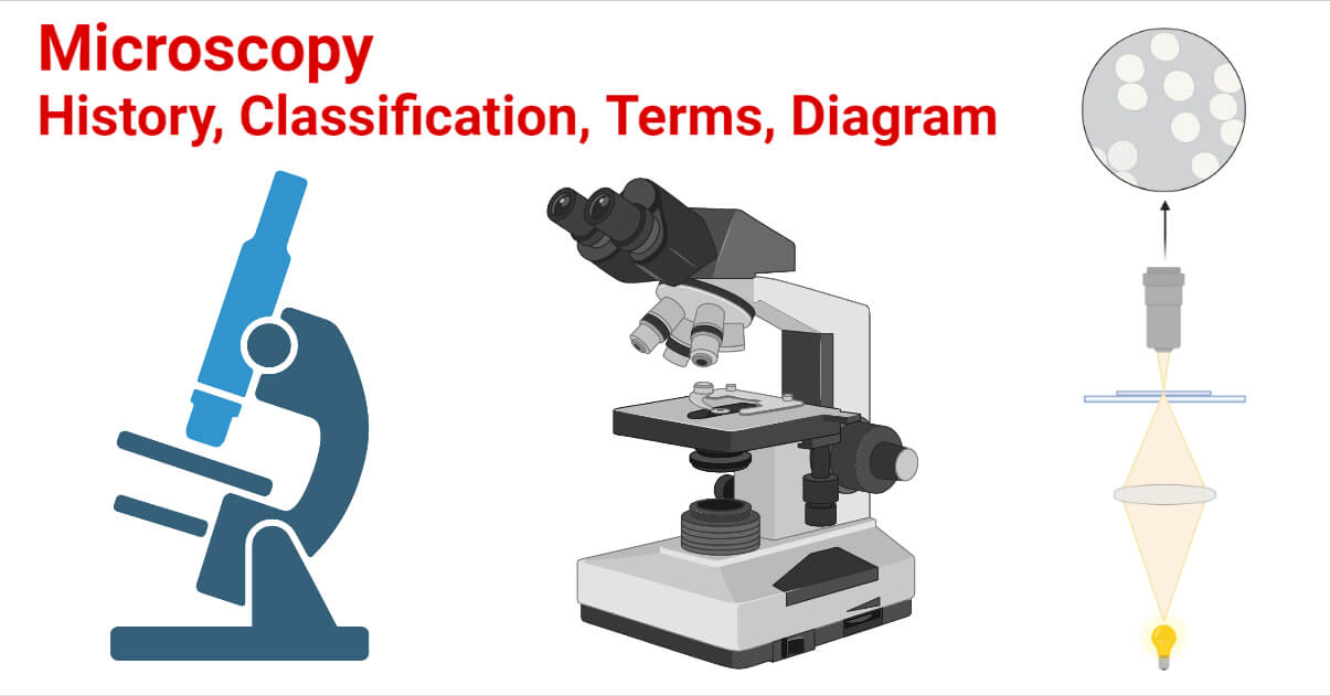
Microscopy- History, Classification, Terms, Diagram
Compound Light Microscope Drawing - paintingvalley.com microscope light diagram parts labeled blank microscopic functions microscopy working label unlabeled Compound Light Micro... 1028x747 11 1 Microscope Light Mic... 344x611 7 1 The Microscope - Com... 1256x1333 6 2 Parts Of Compound Mi... 336x528 5 0 Microscopic Drawing ... 888x762 4 0 Microscopic Drawing ... 2123x2614 2 0 Light Compound Micro...

label microscope diagram | Charts | Microscope, Diagram chart ...
Light Microscope- Definition, Principle, Types, Parts, Labeled Diagram ... Brightfield Light Microscope (Compound light microscope) This is the most basic optical Microscope used in microbiology laboratories which produces a dark image against a bright background. Made up of two lenses, it is widely used to view plant and animal cell organelles including some parasites such as Paramecium after staining with basic stains.

Compound Microscope Parts, Functions, and Labeled Diagram ...
› MSA › collectors_cornerRock Key - Mineralogical Society of America Crystals: Crystals are what minerals form when they are free to grow in nature; like the quartz crystal in the first drawing. In rocks, crystals grow up against each other. They cannot grow as the quartz crystal did in open space. Crystals in rocks have straight edges and they very often show flat shiny faces that reflect light like tiny mirrors.

Microscope With Labels Clip Art at Clker.com - vector clip ...
Join LiveJournal WebPassword requirements: 6 to 30 characters long; ASCII characters only (characters found on a standard US keyboard); must contain at least 4 different symbols;
NSW Biology M1IQ1 a. FHI - Introductory Microscopes & Scale ...
Microscopy - Wikipedia WebOptical or light microscopy involves passing visible light transmitted through or reflected from the sample through a single lens or multiple lenses to allow a magnified view of the sample. The resulting image can be detected directly by the eye, imaged on a photographic plate, or captured digitally.The single lens with its attachments, or the system of lenses …
Microscopes
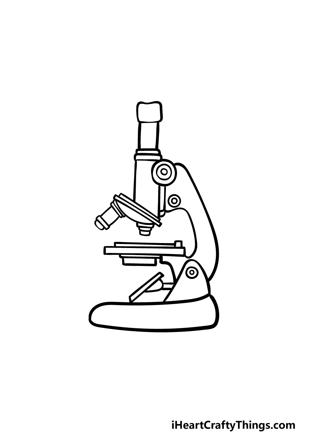
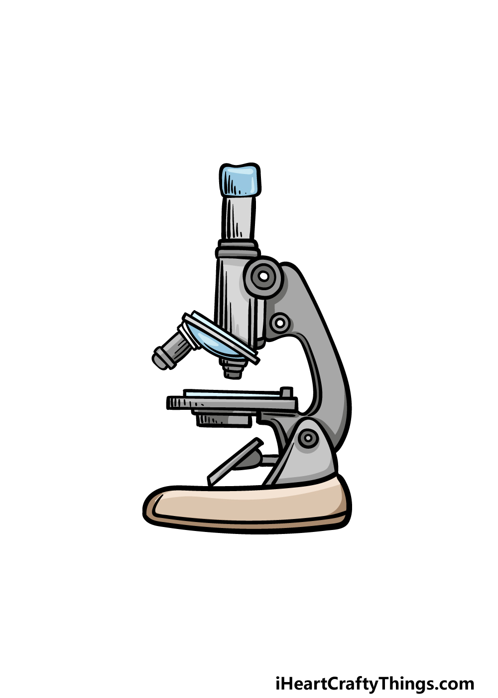
40+ Best Collections Light Microscope Drawing With Label If you are looking for Light microscope drawing with label you've come to the right place. We have collect images about Light microscope drawing with label including images, pictu
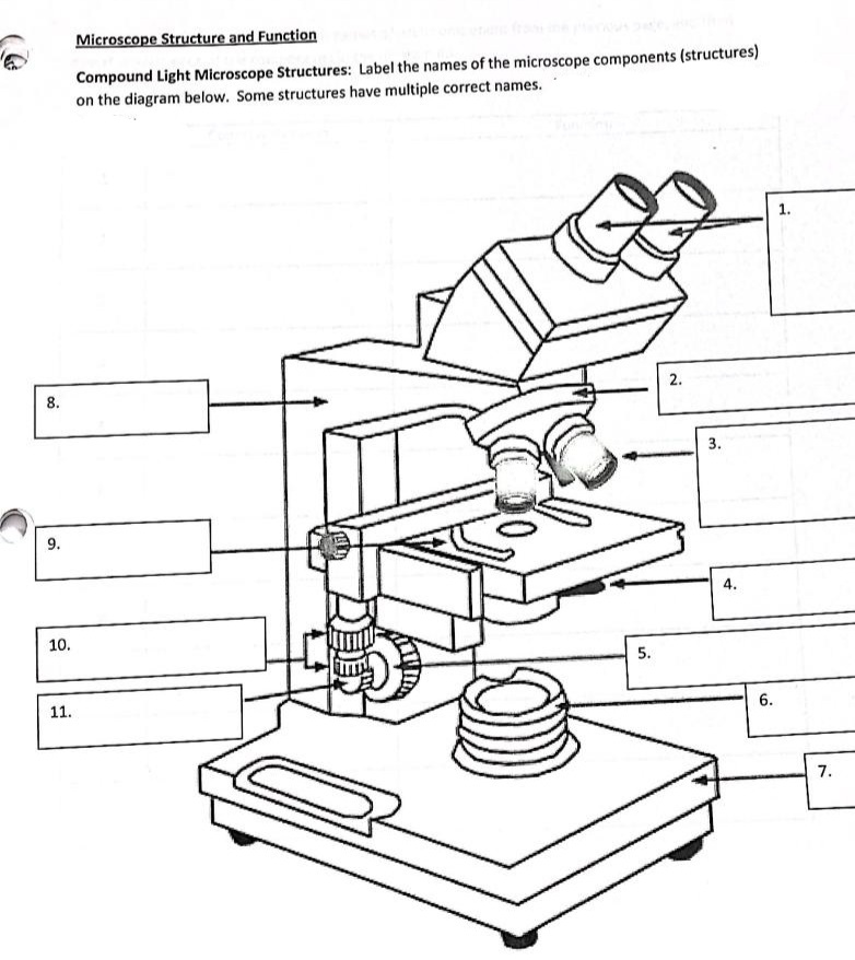
Answered: Microscope Structure and Function… | bartleby
NFL News, Expert Analysis, Rumors, Live Updates, and more - Yahoo! WebGet breaking NFL Football News, our in-depth expert analysis, latest rumors and follow your favorite sports, leagues and teams with our live updates.
Free Microscope Drawing, Download Free Microscope Drawing png ...
How to Sketch a Microscope Slide - Identifying and Sketching Cell ... For a pencil sketch, separate areas into white, light, medium and dark grey and black. To see the light/dark areas, squint so that the hard edges are blurred and your focus is on the shading. Start shading the light areas by following the shapes. For example, shade vertical lines for a flat surface and curved lines for a rounded.

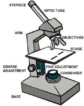
Parts of a Microscope - SmartSchool Systems
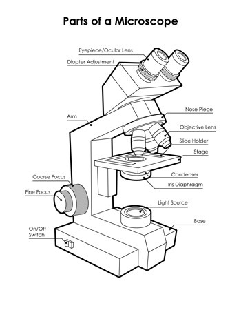
Microscope Parts and Functions Microscope Parts and Functions With Labeled Diagram and Functions How does a Compound Microscope Work? Before exploring microscope parts and functions, you should probably understand that the compound light microscope is more complicated than just a microscope with more than one lens. 5 Hobby Microscopes for Beginners

Compound Light Microscope Labels 1 (Champagne) Diagram | Quizlet
Microscope Diagram Labeled, Unlabeled and Blank | Parts of a Microscope ... Mar 28, 2016 - Print a microscope diagram, microscope worksheet, or practice microscope quiz in order to learn all the parts of a microscope.
Cytology. Cytology. radiation used to illuminate the specimen ...
How to draw compound of Microscope easily - step by step How to draw compound of Microscope easily - step by step - YouTube 0:00 / 6:16 How to draw compound of Microscope easily - step by step Perhaps Bidesh 50.1K subscribers Subscribe 1.3M views 3...

How to draw Microscope diagram for beginners - step by step
Simple Squamous Epithelium under a Microscope with a Labeled Diagram ... Simple squamous epithelium under microscope labeled in renal corpuscle The cortex of a kidney consists of renal corpuscles and the convoluted tubule, straight tubules, nephrons, connecting tubules, and collecting ducts. You will find the medullary ray in the medulla of the kidney that comprises straight tubules and collecting ducts.
Free Microscope Drawing, Download Free Microscope Drawing png ...
A Study of the Microscope and its Functions With a Labeled Diagram ... To better understand the structure and function of a microscope, we need to take a look at the labeled microscope diagrams of the compound and electron microscope. These diagrams clearly explain the functioning of the microscopes along with their respective parts. Man's curiosity has led to great inventions. The microscope is one of them.

Course: s4: Biology , Topic: UNIT 3: MICROSCOPY
No Longer Available - WMUR WebPostal carrier, deputies rally after boy asks Santa for toys and to not get bullied

HOW TO DRAW MICROSCOPE
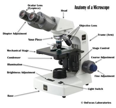
Microscope, Microscope Parts, Labeled Diagram, and Functions Illuminator: Illuminator is the most important microscope parts and it serve as light source for a microscope during slide specimen visualization. It is a continuous source of light (110 volts) used in place of a mirror. The mirror of microscope is used to reflect light from the external light source up through the bottom of the stage.

Microscope - Label - Part 2 Diagram | Quizlet
Patent Public Search | USPTO WebWelcome to Patent Public Search. The Patent Public Search tool is a new web-based patent search application that will replace internal legacy search tools PubEast and PubWest and external legacy search tools PatFT and AppFT.
Labeled Microscope Diagram - Tim's Printables
sports.yahoo.com › nfl › newsNFL News, Expert Analysis, Rumors, Live Updates, and more ... Get breaking NFL Football News, our in-depth expert analysis, latest rumors and follow your favorite sports, leagues and teams with our live updates.

How to draw and label the parts of a microscope? What are at ...
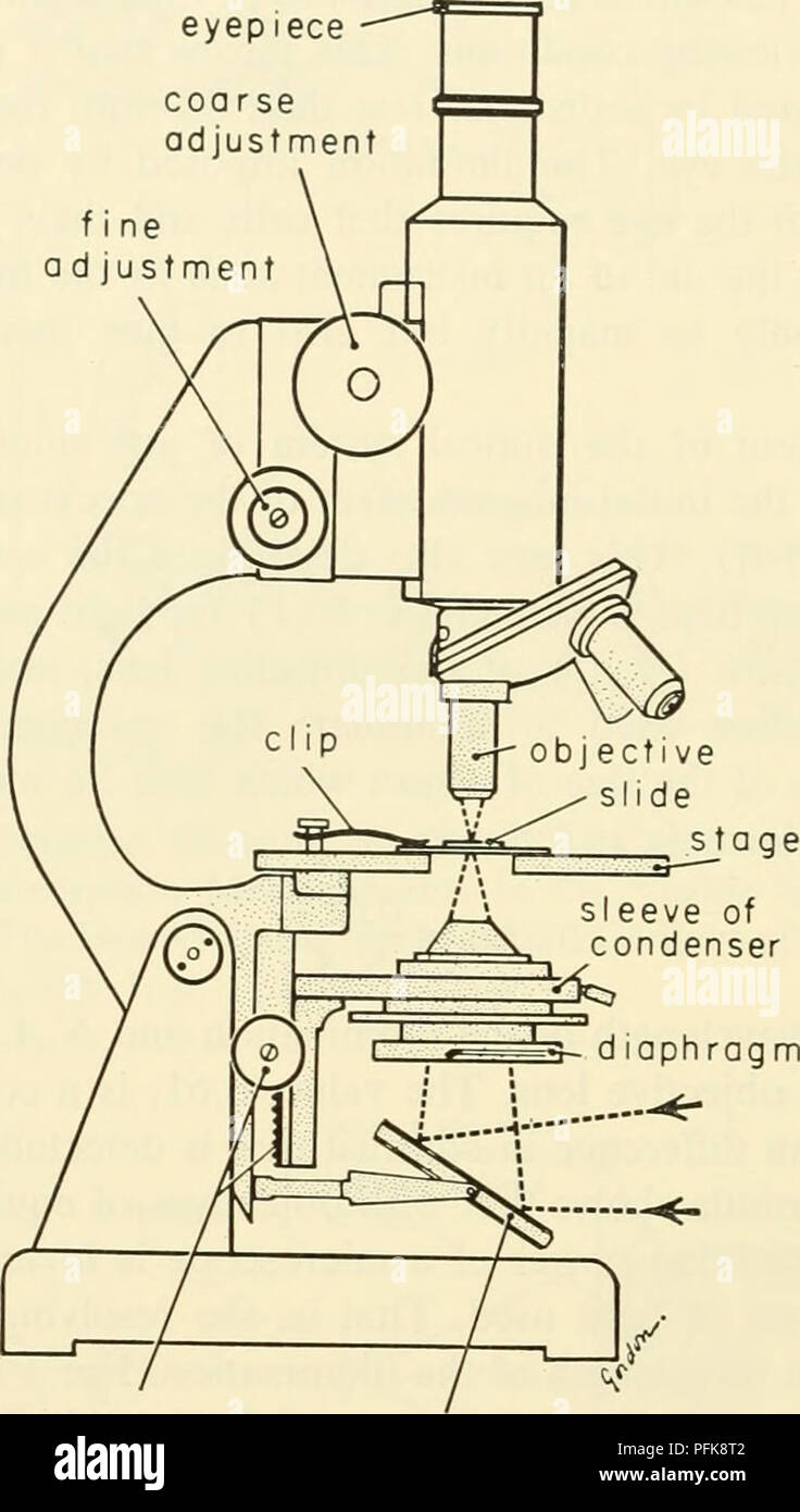
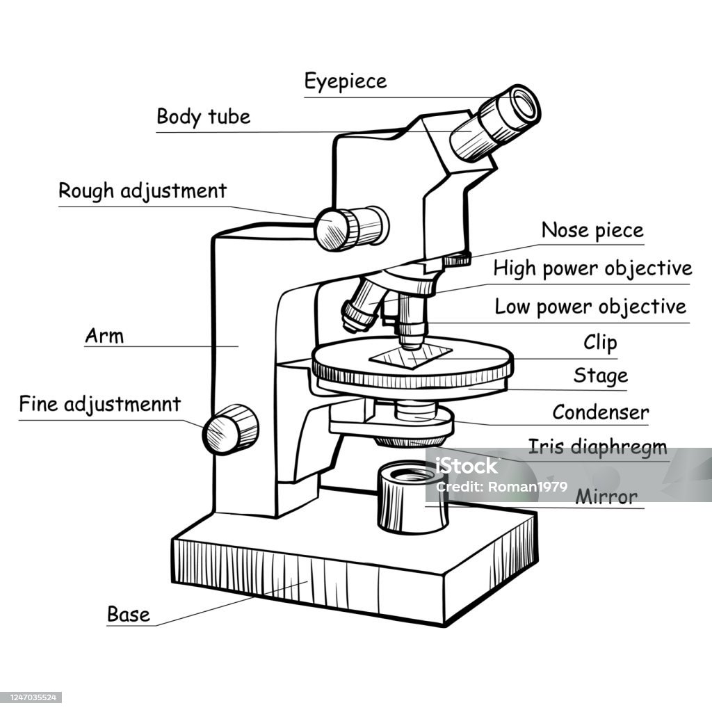
Labelled Diagram of Compound Microscope The below mentioned article provides a labelled diagram of compound microscope. Part # 1. The Stand: The stand is made up of a heavy foot which carries a curved inclinable limb or arm bearing the body tube. The foot is generally horse shoe-shaped structure (Fig. 2) which rests on table top or any other surface on which the microscope in kept.

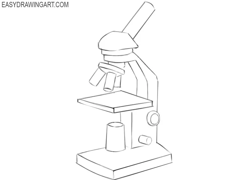
How to Draw a Microscope - Really Easy Drawing Tutorial
Student's Guide: How to Use a Light Microscope Step 1: Connect the light microscope to a power source. If your microscope uses a mirror instead of an illuminator, you can skip this step. Instead, find a place where natural light is easily accessible; Step 2: Turn the revolving nosepiece so the lowest objective lens is in position. Step 3: Mount your specimen onto the stage. But before doing ...
Microscope Clipart Experiment - Simple Drawing Of A ...
Microscope Labeling - The Biology Corner Microscope Labeling. Shannan Muskopf May 31, 2018. This simple worksheet pairs with a lesson on the light microscope, where beginning biology students learn the parts of the light microscope and the steps needed to focus a slide under high power. The labeling worksheet could be used as a quiz or as part of direct instruction where students ...
How to Draw a Microscope - Easy Drawing Art
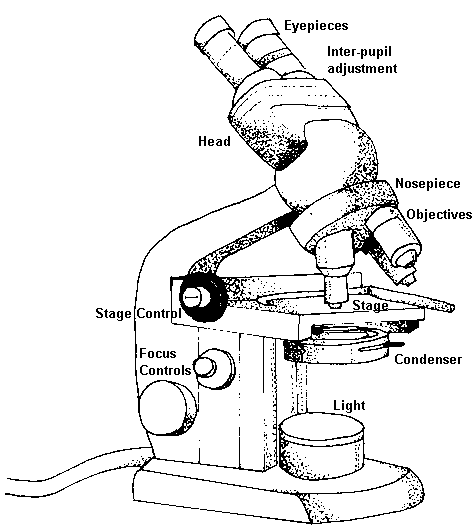
Parts of the Microscope with Labeling (also Free Printouts) A microscope is one of the invaluable tools in the laboratory setting. It is used to observe things that cannot be seen by the naked eye. Table of Contents 1. Eyepiece 2. Body tube/Head 3. Turret/Nose piece 4. Objective lenses 5. Knobs (fine and coarse) 6. Stage and stage clips 7. Aperture 9. Condenser 10. Condenser focus knob 11. Iris diaphragm

13 parts of the Compound Light Microscope Diagram | Quizlet
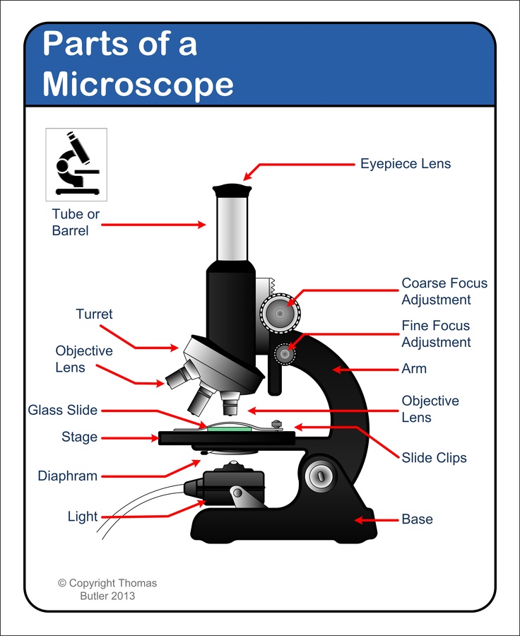
label microscope diagram | Charts | Microscope, Diagram chart, Anatomy ... Microscope parts include eyepiece (10x), objective lenses (4x, 10x, 40x, 100x), fine and coarse focus, slide holder, condenser, iris diaphragm, illuminator, and specimen stage. Without a microscope, we are limited to what we can see with the naked eye.

Labeling the Parts of the Microscope | Microscope World Resources
Microscope Parts, Function, & Labeled Diagram - slidingmotion Microscope parts labeled diagram gives us all the information about its parts and their position in the microscope. Microscope Parts Labeled Diagram The principle of the Microscope gives you an exact reason to use it. It works on the 3 principles. Magnification Resolving Power Numerical Aperture. Parts of Microscope Head Base Arm Eyepiece Lens

Parts of a Microscope | Labeling activities, Science ...
ppubs.uspto.gov › pubwebapp › staticPatent Public Search | USPTO Welcome to Patent Public Search. The Patent Public Search tool is a new web-based patent search application that will replace internal legacy search tools PubEast and PubWest and external legacy search tools PatFT and AppFT.

Simple Microscope - Diagram (Parts labelled), Principle ...
How to Draw a Microscope - Really Easy Drawing Tutorial Easy Microscope Drawing - Step 2 2. Extend a pair of straight, parallel lines from the head, and connect them at the end using a short curved line. This forms the eyepiece tube. The tip of the tube is called the ocular. It is the lens through which you look to view your tiny objects. Easy Microscope Drawing - Step 3 3.

Microscope Drawing Set stock vector. Illustration of sketch ...
Givenchy official site WebDiscover all the collections by Givenchy for women, men & kids and browse the maison's history and heritage

Compound Microscope Parts – Labeled Diagram and their ...
Label the microscope — Science Learning Hub Label the microscope Interactive Add to collection Use this interactive to identify and label the main parts of a microscope. Drag and drop the text labels onto the microscope diagram. eye piece lens diaphragm or iris coarse focus adjustment stage base fine focus adjustment light source high-power objective Download Exercise Tweet

Microscope Diagram Labeled, Unlabeled and Blank | Parts of a ...
› cells › cell_modelInteractive Eukaryotic Cell Model - CELLS alive Cytosol: The cytosol is the "soup" within which all the other cell organelles reside and where most of the cellular metabolism occurs.Though mostly water, the cytosol is full of proteins that control cell metabolism including signal transduction pathways, glycolysis, intracellular receptors, and transcription factors.

Microscope Parts & Functions - AmScope

How to Draw a Microscope - VERY EASY

Compound Microscope: Know Definition,working, diagram, properties

Light Microscope- Definition, Principle, Types, Parts ...
Microscope Drawing - How To Draw A Microscope Step By Step
Microscope Drawing - How To Draw A Microscope Step By Step
Free Microscope Drawing, Download Free Microscope Drawing png ...

microscopy how a microscope works magnification calculations ...

Microscope Diagram Labeled, Unlabeled and Blank | Parts of a ...
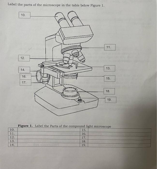
Solved Label the parts of the microscope in the table below ...
Microscope Parts and Functions
Infografis Komponen Mikroskop Lab Elektronik Modern Yang Kuat ...
Post a Comment for "40 light microscope drawing with label"