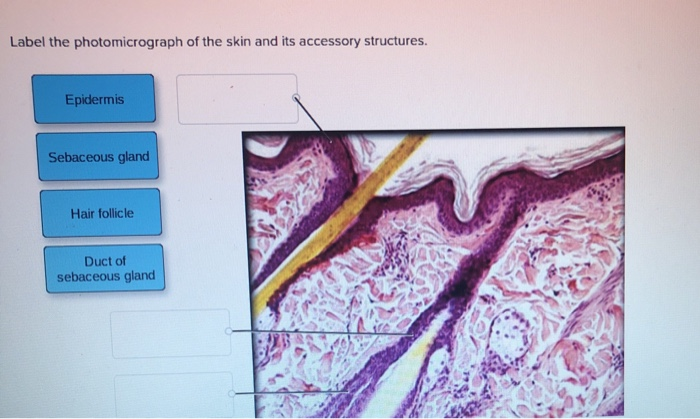
41 label the photomicrograph of the skin and its accessory structures
Label the photomicrograph in Figure 7.4. Examine a slide of hairy skin ... Label the layers of the skin on the diagram and the photograph. Be able to identify the layers on a microscope slide. Look at the skin slide under a microscope. a) Epidermis 1) Stratum corneum ii) Stratum lucidum 111) Stratum granulosum iv) Stratum... Posted one year ago Recent Questions in Basics of Statistics Q: The Skin (Human Anatomy): Picture, Definition, Function, and Skin ... The skin is the largest organ of the body, with a total area of about 20 square feet. The skin protects us from microbes and the elements, helps regulate body temperature, and permits the ...
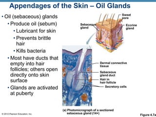
PDF CHAPTER 5 The Integumentary System - LWW tion with its accessory structures. The skin consists of two layers: the epidermis, which includes the outer protective stratum of keratinized epithelial cells, and the dermis, the underlying layer of connective tissue containing blood vessels and nerve endings. The accessory structures include outgrowths of the epidermis (hair and nails).
Label the photomicrograph of the skin and its accessory structures
Anatomy of the Epidermis with Pictures - Verywell Health Summary. The epidermis is composed of layers of skin cells called keratinocytes. Your skin has four layers of skin cells in the epidermis and an additional fifth layer in areas of thick skin. The four layers of cells, beginning at the bottom, are the stratum basale, stratum spinosum, stratum granulosum, and stratum corneum. (Solved) - Label The Photomicrograph Of The Skin And Its Accessory ... Sebaceous gill: The sebum ... LibGuides: BIO 140 - Human Biology I - Textbook: Chapter 10 ... Chapter 15 - Accessory Structures of the Skin ; Section 5 - Digestive System Toggle Dropdown. Chapter 16 - Digestive System Processes and Regulation ; ... The structure of a tissue usually is optimized for its function. Describe how the structure of individual cells and tissue arrangement of the intestine lining matches its main function, to ...
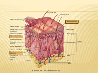
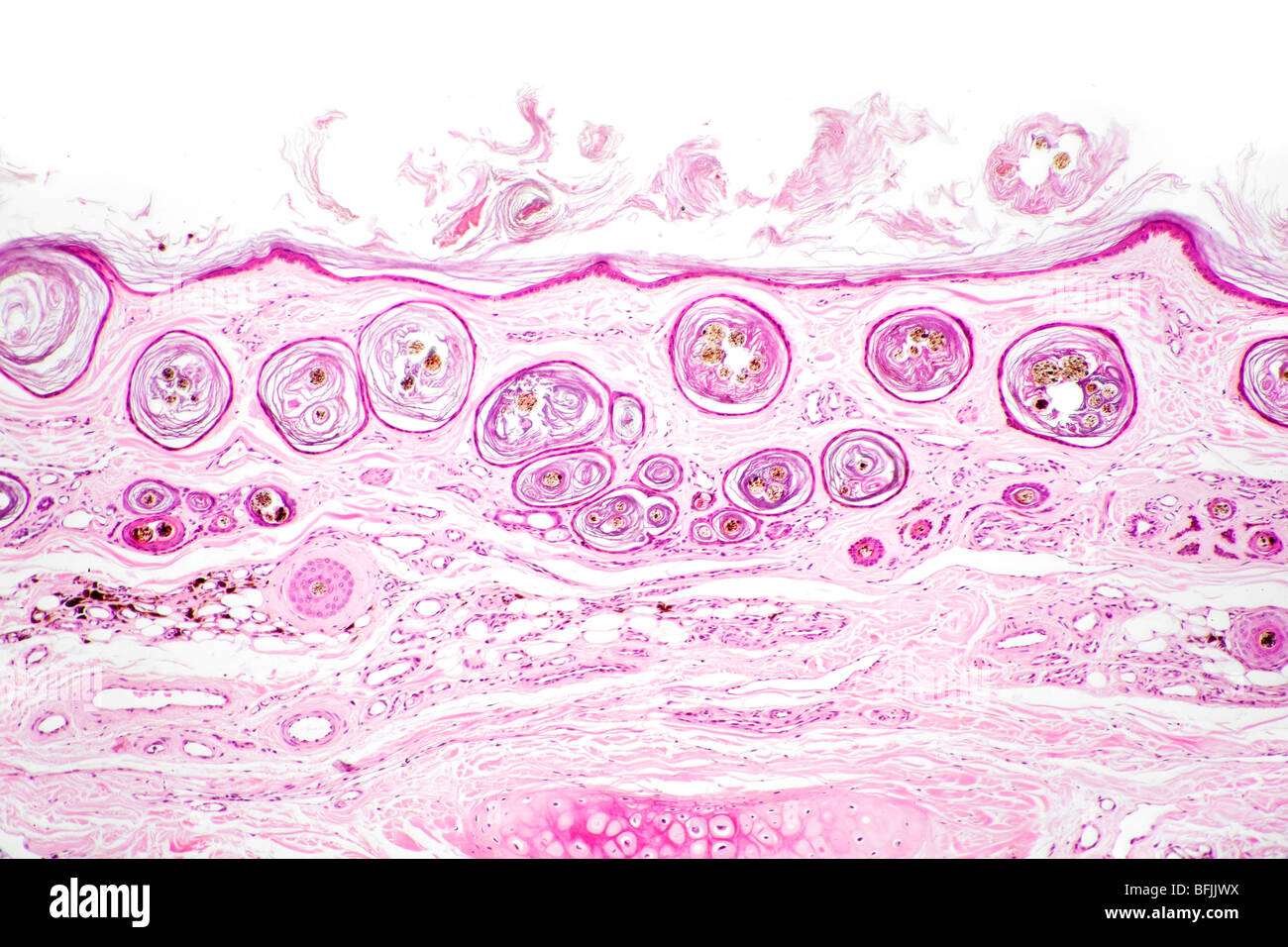
Label the photomicrograph of the skin and its accessory structures. Diagram of human skin structure — Science Learning Hub Diagram of human skin structure. Image. Add to collection. Tweet. Rights: University of Waikato Published 1 February 2011 Size: 100 KB Referencing Hub media. The epidermis is a tough coating formed from overlapping layers of dead skin cells. PreLab03a Integument & Prelab03b Integument Histology Label the photomicrograph of the skin and its accessory structures. epidermis hair follicle duct of sebaceous gland sebaceous gland Recommended textbook solutions Introduction to Anatomy and Physiology 1st Edition Michelle Provost-Craig, Susan J. Hall, William C. Rose 1,678 solutions Hole's Human Anatomy and Physiology unit 4 lab.docx - LAB Unit 4 EXERCISE 7: The Integumentary... FIGURE 7.4:Diagram of the skin and accessory structures. • apocrine (AP-oh-krin) sweat gland • arrector pili (PIE-lee) muscle • eccrine (EK-rin) sweat gland • hair bulb • hair follicle • hair root • hair shaft • papilla (puh-PILL-uh) of hair • sebaceous (se-BAY-shus) gland 1. Hair shaft 2. Hair root 3. Sebaceous glands 4. Arrector pili muscle 5. DiFiore's Atlas of Histology with Functional Correlations (11th Ed.) There is shortage of references in higher teaching institutions especially in newly opened institutions engaged in training of various Veterinary professionals in the country.
Accessory Structures of the Skin | Anatomy and Physiology I - Course Hero Accessory structures of the skin include hair, nails, sweat glands, and sebaceous glands. These structures embryologically originate from the epidermis and can extend down through the dermis into the hypodermis. Hair Figure 1. Hair follicles originate in the epidermis and have many different parts. Diagram of the skin and accessory structures Flashcards | Quizlet Hair shaft. Hair root. Sebaceous gland. Arrector pili muscle. Hair follicle. Hair bulb. Eccrine sweat gland. Papilla of hair. Apocrine sweat gland. Answered: 1. In the photomicrograph below of… | bartleby Science Anatomy and Physiology Q&A Library 1. In the photomicrograph below of cartilage tissue, find and label the indicated structures. Extra cellular r Lacuna Chondrocyte Dyte Elastic protein fibers Extracellular matrix In the photomicrograph below of compact bone tissue, find and label the indicated structu p Osteon Lamella Lacuna o Osteocyte Canaliculi Central canal Kamus Kedokteran Dorland Edisi 31 [z0x292o9mwqn] [Yun.] lebih kecil. rriosis mikros fYrn.l kecil. Juga menunjukkan fraksi dalam sistem metrik (satu per seiuta). photomicrograph, microgram mille lL.l seribu. Juga menunjukkan fraksi dalam sistem metrik. Cf. kilo-. milligram, tnillipede mimetikos [Yun.] meniru. sympalhomimetic nisos [Yun.l kebencian. rzisogamy Lihat -mittent. intromlssion
Figure 7.1: Photomicrograph of Skin Diagram | Quizlet Start studying Figure 7.1: Photomicrograph of Skin. Learn vocabulary, terms, and more with flashcards, games, and other study tools. Solved Label the Photomicrograph of the skin and its - Chegg Expert Answer. 100% (3 ratings) Transcribed image text: Label the Photomicrograph of the skin and its accessory structures. Solved Label the photomicrograph of the skin and its - Chegg Question: Label the photomicrograph of the skin and its accessory structures. Sebaceous gland Duct of sebaceous gland Epidermis Hair follicle This problem has been solved! See the answer Show transcribed image text Expert Answer 100% (19 ratings) Layers of the Skin | Anatomy and Physiology I | | Course Hero The skin and its accessory structures make up the integumentary system, which provides the body with overall protection. The skin is made of multiple layers of cells and tissues, which are held to underlying structures by connective tissue (Figure 1). The deeper layer of skin is well vascularized (has numerous blood vessels).
1 - Clinical Significance of Dental Anatomy, Histology ...
Skin Layers: Structure, Function, Anatomy, and More - Verywell Health Collagen is the main protein that provides structure to skin and connective tissues. It gives skin its elasticity and strength. The thickness of the dermis varies by its location on the body. On the eyelids, it is roughly 0.6 millimeters thick. On the back, palms of hands, and soles of the feet, it's 3 millimeters thick.

Hair, Hair Follicles, Sebaceous Glands, and Sudoriferous ...
Do You Know Your Skin? Layers of the Epidermis and their Functions Stratum Corneum. This is the outermost layer of the epidermis that insulates the skin from the outside environment. It acts like a protective covering, keeping the moisture trapped inside the skin. Basically, the layer seals the skin keeping its contents intact. It is due to this layer that the skin is impermeable to quite a few chemicals and ...
Skin histology hi-res stock photography and images - Alamy
photomicrograph of thick skin Diagram | Quizlet skin structure (part 2 of 2) 8 terms. mckennawebber. nail structure (part 1 of 2) 8 terms. mckennawebber. Other sets by this creator. selection of exercise type and parameters. ... Start studying photomicrograph of thick skin. Learn vocabulary, terms, and more with flashcards, games, and other study tools.

Fe2+: Fe3+ Molar Ratio Influences the Immunomodulatory ...
Figure 7.4 Photomicrograph of the skin and accessory structures - Quizlet Papilla of hair. a projection of connective tissue into the hair follicle and contains blood vessels that provide nutrients to the dividing cells of the matrix. Sebaceous Gland. Oil glands that surround hair follicles; secrete oils that lubricates skin, hair, and into the neck of the hair follicle. Hair Follicle.

Hair shaft Dermal papillae Epidermis Subpapillary vascular ...
The Integumentary System Lab Report - 703 Words | Studymode A tan is your skin's attempt to prevent UV rays from doing any further damage to the sensitive skin cells in your epidermis. …show more content… Be sure to label all of the structures in the epidermis and dermis you were able to find: QUESTIONS: A. Compare your slide to the photomicrograph example in the lab procedure.

skin and accessory structures diagram Diagram | Quizlet
Anatomy and Physiology Homework Chapter 6 Flashcards | Quizlet Study with Quizlet and memorize flashcards containing terms like Label the parts of the skin and subcutaneous tissue. -Blood Capillaries -Piloerector muscle -Dermal papilla -Hair bulb -Sensory nerve fibers -Tactile corpuscle -Hair follicle -Sebaceous gland, Label the parts of the skin and subcutaneous tissue. -Hypodermis -Sweat pores -Dermis -Hairs -Cutaneous blood vessels -Epidermis -Sweat ...
Skin and the Integumentary System
(PDF) Raven Biology of Plants | Michael Branks - Academia.edu Enter the email address you signed up with and we'll email you a reset link.

Respiratory System - ScienceDirect
Solved Label the photomicrograph of the skin and its - Chegg Science. Anatomy and Physiology. Anatomy and Physiology questions and answers. Label the photomicrograph of the skin and its accessory structures. Epidermis Sebaceous gland Hair follicle Duct of sebaceous gland Label the photomicrograph of the skin and its accessory structures.

Label the photomicrograph of the skin and its accessory ...
PDF The Integumentary System - Holly H. Nash-Rule, PhD Label the skin structures and areas indicated in the accompanying diagram of thin skin. Then, complete the statements that ... Accessory Organs of the Skin 9. Match the key choices with the appropriate descriptions. Some terms are used more than once. Key: a. arrector pili d. hair follicle g.

Stratum Lucidum 5 Translucent layer found in thick skin ...
Solved Label the photomicrograph of thick skin | Chegg.com Anatomy and Physiology questions and answers; Label the photomicrograph of thick skin ; Question: Label the photomicrograph of thick skin . This problem has been solved! See the answer See the answer See the answer done loading. Show transcribed image text Expert Answer. Who are the experts?

Label the photomicrograph of the skin and its accessory ...
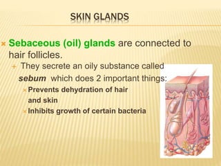
Accessory Structures of the Skin - Anatomy & Physiology Accessory structures of the skin include hair, nails, sweat glands, and sebaceous glands. These structures embryologically originate from the epidermis and can extend down through the dermis into the hypodermis. Hair Hair is a keratinous filament growing out of the epidermis. It is primarily made of dead, keratinized cells.
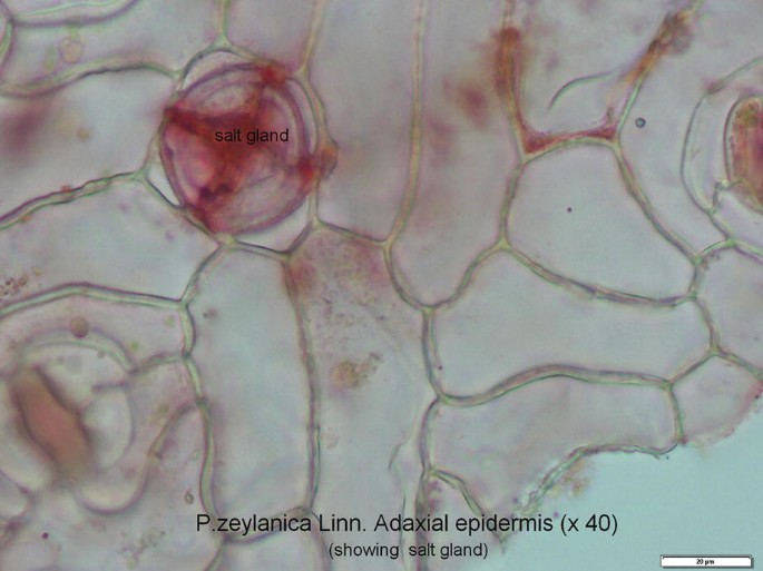
A Comparative Account of the Morphological and Anatomical ...
Structure and Function of Skin | Biology for Majors II - Course Hero The skin and its accessory structures make up the integumentary system, which provides the body with overall protection. The skin is made of multiple layers of cells and tissues, which are held to underlying structures by connective tissue (Figure 1). The deeper layer of skin is well vascularized (has numerous blood vessels).

Photomicrograph of rat seminal vesicles stained with HE ...
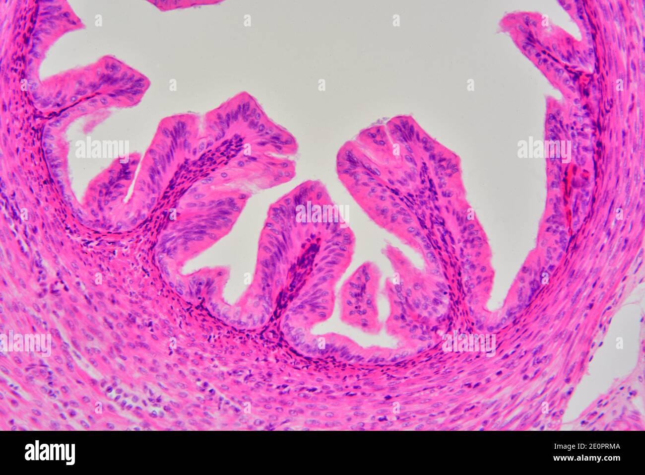
Solved Label the photomicrograph of the skin and its - Chegg Label the photomicrograph of the skin and its accessory structures Epidermis Duct of sebaceous gland Hair follicle Sebaceous gland Show transcribed image text Expert Answer 2. The picture here demonstrates the pseudostratified columnar epithelium.
Handout: The Integumentary System Anatomy & Physiology I ...
PDF Name the Condition the cartoon and the photomicrograph. •Name the Layers of skin and label the dermal papilla and dermis •Name the Layers of skin and label the dermal papilla and dermis. Name the layer of skin shown. Stratum Spinosum. Name the specific layers of skin indicated by the ... Identify the following structures: Epidermis, Hair cortex, Hair medulla ...
Integumentary System – Building a Medical Terminology Foundation
RS7 (2).pdf - Basic Structure of the Skin 1. Complete the... Label the integumentary structures and areas indicated in the diagram. ( Labeled from bottom right to bottom left) Sweat glands Dermis Epidermis Hair shaft Sebaceous gland Hair follicle Hair root Arrector pili muscle Dermis Hair root Arrector pili muscle Dermis 5.Label the layers of the epidermis in thick skin.
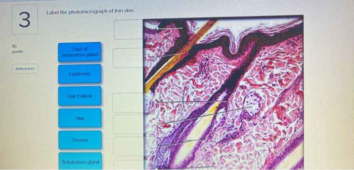
Solved Label the photomicrograph of thin skin 3 10 points ...
LibGuides: BIO 140 - Human Biology I - Textbook: Chapter 10 ... Chapter 15 - Accessory Structures of the Skin ; Section 5 - Digestive System Toggle Dropdown. Chapter 16 - Digestive System Processes and Regulation ; ... The structure of a tissue usually is optimized for its function. Describe how the structure of individual cells and tissue arrangement of the intestine lining matches its main function, to ...
Integumentary System Overview
(Solved) - Label The Photomicrograph Of The Skin And Its Accessory ... Sebaceous gill: The sebum ...

Figure 7.4 Photomicrograph of the skin and accessory ...
Anatomy of the Epidermis with Pictures - Verywell Health Summary. The epidermis is composed of layers of skin cells called keratinocytes. Your skin has four layers of skin cells in the epidermis and an additional fifth layer in areas of thick skin. The four layers of cells, beginning at the bottom, are the stratum basale, stratum spinosum, stratum granulosum, and stratum corneum.
Chapter 5
Integumentary System Overview
Chapter 5
Lamina propria hi-res stock photography and images - Alamy

Structure and Function of Skin | Biology for Majors II

5.1 Layers of the Skin – Anatomy & Physiology
A Comparative Account of the Morphological and Anatomical ...
Macroscopic and microscopic morphology of the trachea and ...

Brief exposure of neonatal testis cells to EGF or GDNF alters ...
Patterns of heterogeneous expression of pannexin 1 and ...
Nutrients | Free Full-Text | Amelioration of Ethanol-Induced ...
Frontiers | Relaxin-3 Innervation From the Nucleus Incertus ...

Photomicrograph of rat testes stained with HE (× 200, scale ...

A&P 139 Chapter 22 Reproductive System Flashcards | Quizlet
Solved Label the photomicrograph of the skin and its | Chegg.com
Skin cross section hi-res stock photography and images - Alamy
Ch 4 Skin Power Point

Chapter 15 - Accessory Structures of the Skin - BIO 140 ...
Disruptions / Hypoxic-Ischemic Injury (Section 4) - Perinatal ...

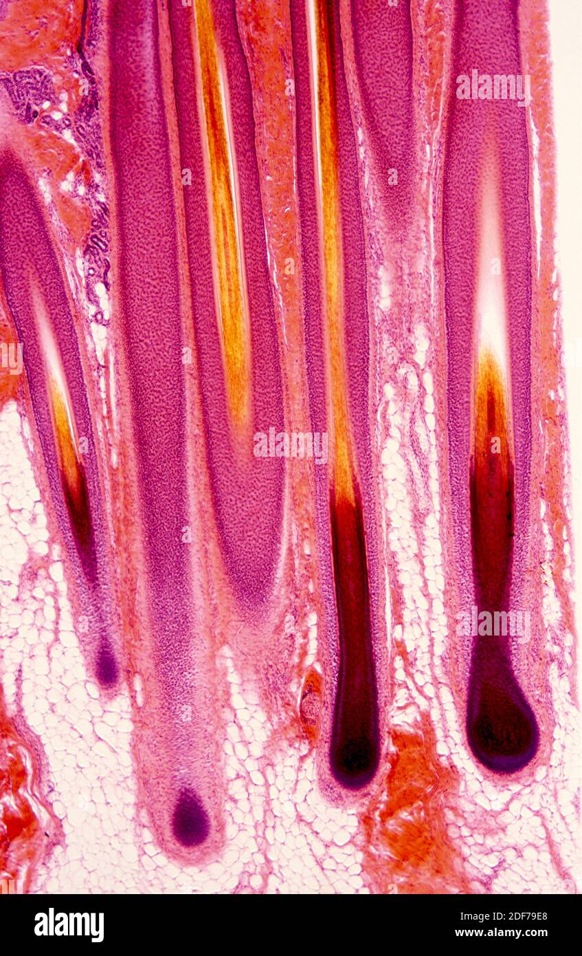
Human scalp, light micrograph - Stock Image - C054/3494 ...
Integumentary System Overview
Novel quantitative signature of tumor stromal architecture ...
Chapter 5
Post a Comment for "41 label the photomicrograph of the skin and its accessory structures"