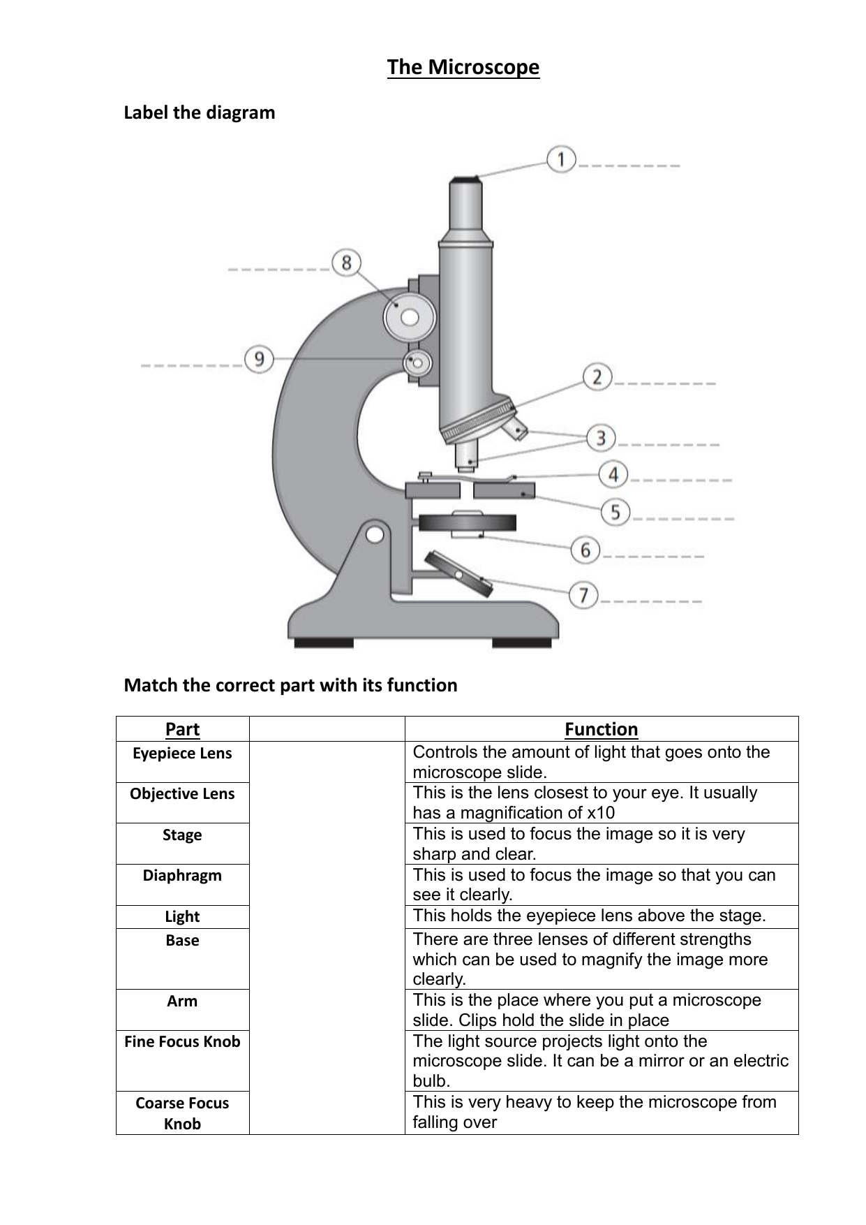
40 label and function of microscope
Microscope labeling and functions Flashcards | Quizlet Microscope labeling and functions. STUDY. Flashcards. Learn. Write. Spell. Test. PLAY. Match. Gravity. Created by. mveet. Terms in this set (27) Separates the eyepiece lens from the objective lenses. Body Tube. Holds the low-power and high-power objective lenses; allows the lenses to rotate for viewing. Microscope Parts and Functions Flashcards | Quizlet Eyepiece. Used to view an object under the microscope, has a magnification power of 10X. Nosepiece. Holds the objective lenses and rotates to change the magnification. Scanning Lens. Magnifies the image 4x and is found on the nosepiece. Medium Power objective. Magnifies the image 10x and is found on the nosepiece. Slide.
A Study of the Microscope and its Functions With a Labeled Diagram The camera present within the microscope captures images to reveal the finer details of the specimen. This microscope can zoom and view the density of a specimen until it is only a micrometer thick and has a magnification ranging between 1,000 - 250,000x on the fluorescent screen. This microscope needs a computer software to yield precise results.

Label and function of microscope
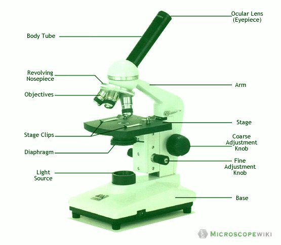
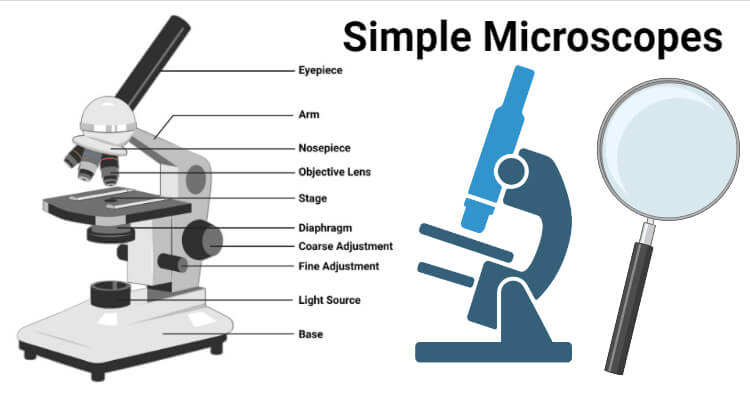
Parts of the Microscope with Labeling (also Free Printouts) Let us take a look at the different parts of microscopes and their respective functions. 1. Eyepiece it is the topmost part of the microscope. Through the eyepiece, you can visualize the object being studied. Its magnification capacity ranges between 10 and 15 times. 2. Body tube/Head It is the structure that connects the eyepiece to the lenses. Solved Label the diagram below to compare the structure and - Chegg Label the diagram below to compare the structure and function of a light microscope versus an electron microscope. Light Microscope Transmission Electron Microscope Viewing screen Objective lens Electron gun Image Condenser lens Glass Electromagnet Electron beams Light rays Ocular lens Specimen Eye Electromagnet Glass Lamp Glass Electromagnet Zoom label the parts of a microscope - TeachersPayTeachers Check out this well-organized Microscope label and describe worksheet. Part I is a visual that students will label and it corresponds with Part II where they will describe the function of those parts. This is a great worksheet that can easily be used for classwork, group work, homework, assessments .
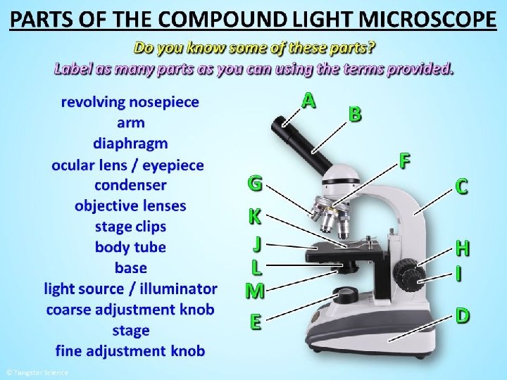
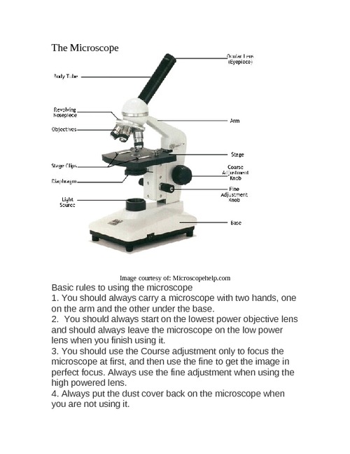
Label and function of microscope. Labeling the Parts of the Microscope Labeling the Parts of the Microscope This activity has been designed for use in homes and schools. Each microscope layout (both blank and the version with answers) are available as PDF downloads. You can view a more in-depth review of each part of the microscope here. Download the Label the Parts of the Microscope PDF printable version here. Parts of a Microscope - The Comprehensive Guide Step 1: Fully open field and condenser diaphragms and focus on specimen using x10 objective. Step 2: Fully close field diaphragm and adjust the condenser and focus so edges are as sharp as possible. Step 3: Use screws at front of condenser to centre field diaphragm and open field diaphragm to fill view. Step 4: Remove eyepiece and close down ... Microscope- Definition, Parts, Functions, Types, Diagram, Uses A microscope is an optical instrument having one or more lenses system which is used to get a clear magnified image of minute objects or structures that can't be viewed by the naked eyes. Derived from Greek words "mikrós " meaning "small" and "skópéō" meaning "look at " . They are devices used to observe the detailed structure of small objects. Label the microscope — Science Learning Hub All microscopes share features in common. In this interactive, you can label the different parts of a microscope. Use this with the Microscope parts activity to help students identify and label the main parts of a microscope and then describe their functions. Drag and drop the text labels onto the microscope diagram.
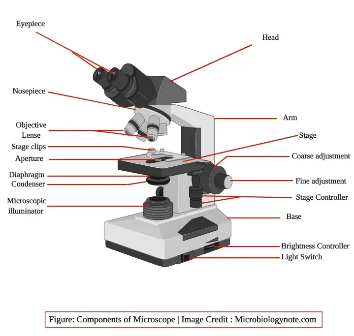
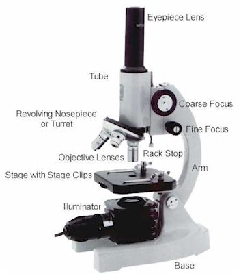
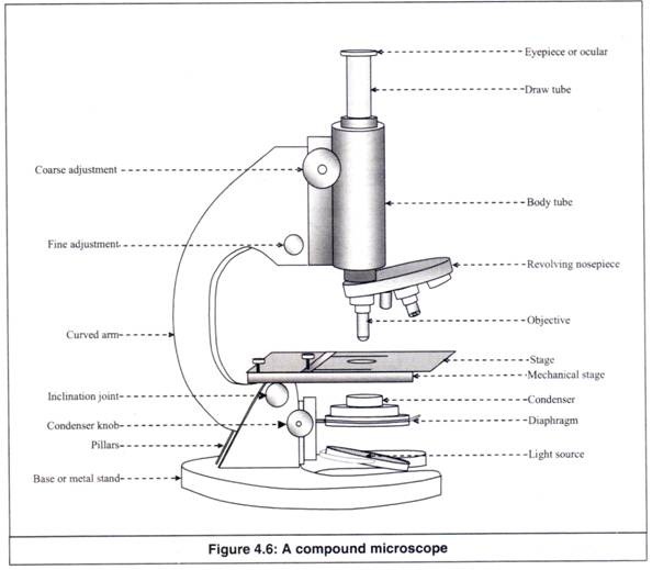
Parts of a Compound Microscope (And their Functions) List of Microscope Parts and their Functions. 1. Ocular Tubes (Monocular, Binocular & Trinocular) The ocular tubes, are to tubes that lead from the head of the microscope out to your eyes. On the end of the ocular tubes are usually interchangeable eyepieces (commonly 10X and 20X) that increase magnification. Compound Microscope: Definition, Diagram, Parts, Uses, Working ... - BYJUS The compound microscope is mainly used for studying the structural details of cell, tissue, or sections of organs. The parts of a compound microscope can be classified into two: Non-optical parts Optical parts Non-optical parts Base The base is also known as the foot which is either U or horseshoe-shaped. Parts of a Compound Microscope and Their Functions It controls the amount and intensity of light that enters the microscope. It can be either an iris diaphragm or a disc diaphragm. Condenser: It's a lense that's hidden beneath the stage. The size of the light beam is controlled by it. It collects and directs light from the mirror to the objective lens. Microscope Parts, Function, & Labeled Diagram - slidingmotion Microscope Parts Labeled Diagram The principle of the Microscope gives you an exact reason to use it. It works on the 3 principles. Magnification Resolving Power Numerical Aperture. Parts of Microscope Head Base Arm Eyepiece Lens Eyepiece Tube Objective Lenses Nose Piece Adjustment Knobs Stage Aperture Microscopic Illuminator Condenser Lens
Microscope parts — Science Learning Hub Microscope parts — Science Learning Hub Microscope parts Add to collection In this activity, students identify and label the main parts of a microscope and describe their function. By the end of this activity, students should be able to: identify the main parts of a microscope describe the function of the different parts of a microscope. Simple Microscope - Parts, Functions, Diagram and Labelling Simple Microscope - Parts, Functions, Diagram and Labelling A microscope is one of the commonly used equipment in a laboratory setting. A microscope is an optical instrument used to magnify an image of a tiny object; objects that are not visible to the human eyes. Table of Contents The common types of microscopes are: What is a Simple microscope? Microscope Parts and Functions First, the purpose of a microscope is to magnify a small object or to magnify the fine details of a larger object in order to examine minute specimens that cannot be seen by the naked eye. Here are the important compound microscope parts... Eyepiece: The lens the viewer looks through to see the specimen. Microscope Types (with labeled diagrams) and Functions Microscopes aid in identifying viruses, bacteria and other microbial organisms and help the researchers in studying about diseases and find a cure to them. Researchers and pathologists examine several specimens a day to understand the underlying cause to a disease to enable doctors to give the right treatment to their patients
Labeling the Parts of the Microscope | Microscope activity ...
Compound Microscope Parts, Functions, and Labeled Diagram Each part of the compound microscope serves its own unique function, with each being important to the function of the scope as a whole. The individual parts of a compound microscope can vary heavily depending on the configuration & applications that the scope is being used for. Common compound microscope parts include:

Parts of a Compound Microscope and Their Functions
Simple Microscope - Diagram (Parts labelled), Principle, Formula and Uses Simple microscope is a magnification apparatus that uses a combination of double convex lens to form an enlarged, erect image of a specimen. The working principle of a simple microscope is that when a lens is held close to the eye, a virtual, magnified and erect image of a specimen is formed at the least possible distance from which a human eye ...

Microscope Diagram Labeled, Unlabeled and Blank | Parts of a ...
Compound Microscope Parts - Labeled Diagram and their Functions - Rs ... The term "compound" refers to the microscope having more than one lens. Basically, compound microscopes generate magnified images through an aligned pair of the objective lens and the ocular lens. In contrast, "simple microscopes" have only one convex lens and function more like glass magnifiers.

Compound Microscope Parts, Functions, and Labeled Diagram ...
Microscope Parts & Functions - AmScope Invented by a Dutch spectacle maker in the late 16th century, compound light microscopes use two sets of lenses to magnify images for study and observation. The first set of lenses are the oculars, or eyepieces, that the viewer looks into; the second set of lenses are the objectives, which are closest to the specimen.
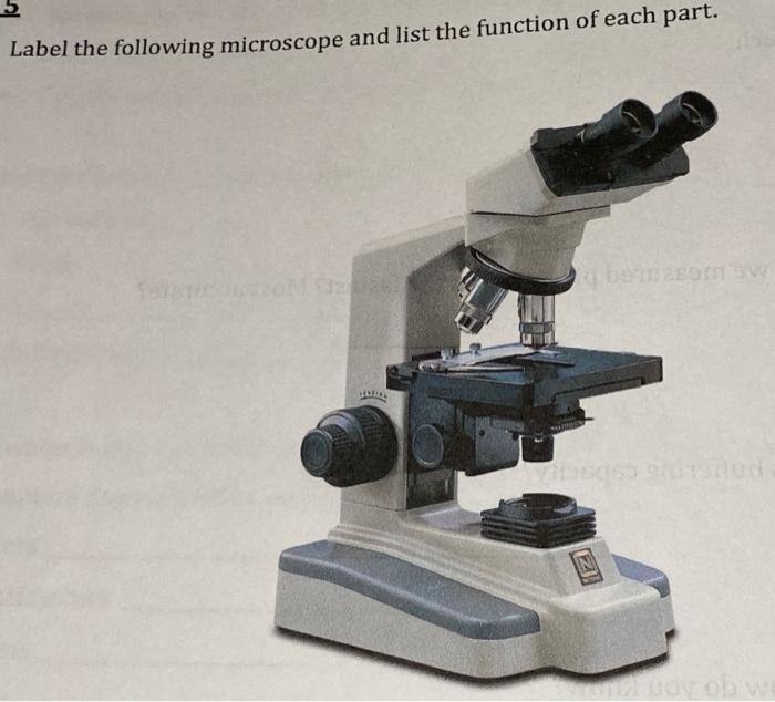
Solved Label the following microscope and list the function ...
Light Microscope- Definition, Principle, Types, Parts, Labeled Diagram ... The functioning of the light microscope is based on its ability to focus a beam of light through a specimen, which is very small and transparent, to produce an image. The image is then passed through one or two lenses for magnification for viewing. The transparency of the specimen allows easy and quick penetration of light.

Simple Microscope - Diagram (Parts labelled), Principle ...
Label And Describe Parts Of A Microscope Teaching Resources | TpT Check out this well-organized Microscope label and describe worksheet. Part I is a visual that students will label and it corresponds with Part II where they will describe the function of those parts. This is a great worksheet that can easily be used for classwork, group work, homework, assessments and more!

Parts of a Microscope Labeling Activity
Light Microscope: Functions, Parts and How to Use It The function of the light microscope is based on its ability to focus a beam of light through a very small and transparent specimen, to produce an image. The image is then passed through one or two lenses for magnification to view. The transparency of the specimen allows for easy and fast light penetration. Specimens can vary from bacteria to ...

Parts of Microscope, Function, Names & Labeled Diagram ...
Microscope, Microscope Parts, Labeled Diagram, and Functions Microscopes magnify or enlarge small objects such as cells, microbes, bacteria, viruses, microorganisms etc. at a viewable scale for examination and analysis. Microscopes consist of one or more magnification lenses to enlarge the image of the microscopic objects placed in the focal plane.

label microscope diagram | Charts | Microscope, Anatomy bones ...
Types of Microscopes: Definition, Working Principle, Diagram ... A compound microscope is defined as the type of microscope that has more than one lens. It has a combination of lenses and two optical parts known as an objective lens and eyepiece or ocular lens. The magnifying power of the compound microscope is given as: Where, D is the least distance of distinct vision L is the length of the microscope tube

microscope | Types, Parts, History, Diagram, & Facts | Britannica
Microscope Of Parts Function Quiz And Microscope to label some of the worksheets for this concept are label parts of the microscope answers the microscope parts and use label parts of the microscope parts of the light microscope labeling scientific tools microscope name use the word list to help you label the 12 7 3 science light microscope wo rk name period date lab 3 use of the Each part plays a vital role in our hearing ...

List: Parts of a Microscope and their Function | Pathwooded
Parts of a microscope with functions and labeled diagram Microscopes are made up of lenses for magnification, each with its own magnification powers. Depending on the type of lens, it will magnify the specimen according to its focal strength. Their ability to function is because they have been constructed with special components that enable them to achieve high magnification levels.

Parts of a Microscope - SmartSchool Systems
label the parts of a microscope - TeachersPayTeachers Check out this well-organized Microscope label and describe worksheet. Part I is a visual that students will label and it corresponds with Part II where they will describe the function of those parts. This is a great worksheet that can easily be used for classwork, group work, homework, assessments .
Introduction To Medical Technology Lecture 9 Introduction ...
Solved Label the diagram below to compare the structure and - Chegg Label the diagram below to compare the structure and function of a light microscope versus an electron microscope. Light Microscope Transmission Electron Microscope Viewing screen Objective lens Electron gun Image Condenser lens Glass Electromagnet Electron beams Light rays Ocular lens Specimen Eye Electromagnet Glass Lamp Glass Electromagnet Zoom

What is a Compound Microscope? | Microscope World Blog
Parts of the Microscope with Labeling (also Free Printouts) Let us take a look at the different parts of microscopes and their respective functions. 1. Eyepiece it is the topmost part of the microscope. Through the eyepiece, you can visualize the object being studied. Its magnification capacity ranges between 10 and 15 times. 2. Body tube/Head It is the structure that connects the eyepiece to the lenses.

Simple Microscope - Diagram (Parts labelled), Principle ...
parts of a microscope - Clip Art Library
Parts of Microscope with their Functions - Microscope parts

Microscope Diagram and Functions | Science fair projects ...
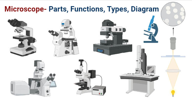
Microscope- Definition, Parts, Functions, Types, Diagram, Uses
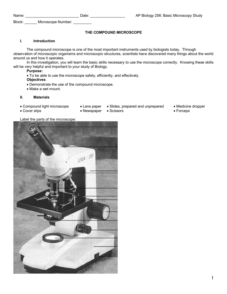
AP Introduction Microscopy Lab

Label Microscope Diagram - EnchantedLearning.com
Microscope Parts & Specifications | Microscope World Resources
Working Principle and Parts of a Compound Microscope (with ...

Dissecting Stereo Microscope Parts and Functions

Parts of Dissecting Microscope | Botany

Compound Microscope Parts, Function, & Diagram | What is a ...

Compound Microscope Parts – Labeled Diagram and their ...

Parts of a Microscope - TessShaheenmicroscopy
Microscope Types (with labeled diagrams) and Functions
Types of Microscopes | Microscope World Blog

PARTS OF MICROSCOPE| LEARN TO LABEL COMPOUND MICROSCOPE| JUST ...
Microscope- Definition, Parts, Functions, Types, Diagram, Uses

13 parts of the Compound Light Microscope Diagram | Quizlet

5 Important Types of Microscopes used in Biology (With Diagram)
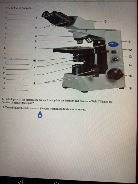
Solved Label all Identified parts. 10 12 13 14 12. 13. 14 ...
Microscope - Teaching resources

Simple Microscope - Parts, Functions, Diagram and Labelling ...

Label the microscope — Science Learning Hub
Simple Microscope- Definition, Principle, Magnification ...
Microscope worksheet 1
Post a Comment for "40 label and function of microscope"